|
|


Tap the top screen.

Tap and select the desired item from the menu screen.
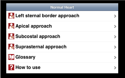

Likewise, tap a sub-item, if any.

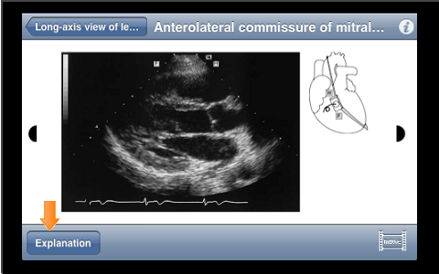
On the still image screen, tap the "Explanation" button at the lower area of the screen to display the explanation screen.
To return to the still image screen, tap the "Image" button.

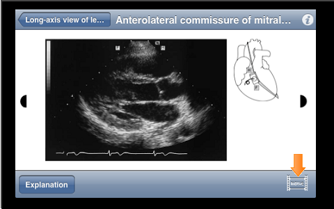
To see a video, tap any one of video link buttons at the lower area of the screen.
You can access videos from either of the still image screen or the explanation screen, as they are linked.

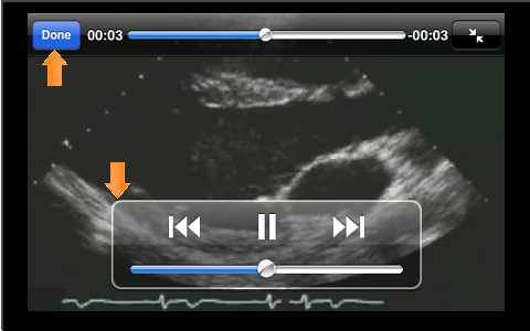
Single-tap on a moving image screen (full-screen) displays the operation panel, where you can playback, fast-forward, and reverse the moving images.
Tap the screen area other than the operation panel again, or leave it for a while without any operation, the operation panel disappears.
Screen returns to the video only display.
Also, to end the playback of video, tap the "End" button at the upper area of the screen so as to return to the previous screen before the playback of video starts (that is, the screen goes back to either of the still image or explanation screen).

- In "#1-Normal Heart", by tapping the i button on the upper right of the screen for each case, the terminology of abbreviations specially used in the case appear. Tap the screen again to return. The terminology contains the abbreviations that are useful in "#1-Normal Heart" only. You can also select abbreviations from the top menu.
- On the still image screen and examination screen, a half-moon-shaped button appears by tapping the right or left upper corner of the page. Tap this button again allows you to access the next case page without returning to the sub-menu screen.
- Zooming by pinch-in/pinch-out (an operation to enlarge or shrink the screen using two fingers) is available on each screen.
Meaning of video icons
- Scanning region
 ; Parasternal region ; Parasternal region
 ; Apical area ; Apical area
 ; Epigastrium ; Epigastrium
 ; Suprasternal space ; Suprasternal space
- Cross sectional image
Long; Long-axis view of left ventricle/Long-axis view of blood vessel
Short; Short-axis view of left ventricle/Short-axis view of blood vessel
RV out; Long-axis view of right ventricular outflow tract
RV in; Long-axis view of right ventricular inflow tract
4 cv; 4-chamber view
2 cv; 2-chamber view
Sagi; Sagittal view
Coro; Coronal view
- Imaging mode
M; M-mode
Color; Color flow imaging
PD; Pulsed Doppler
CWD; Continuous wave Doppler
| ↑ TOP |
|
|
|